

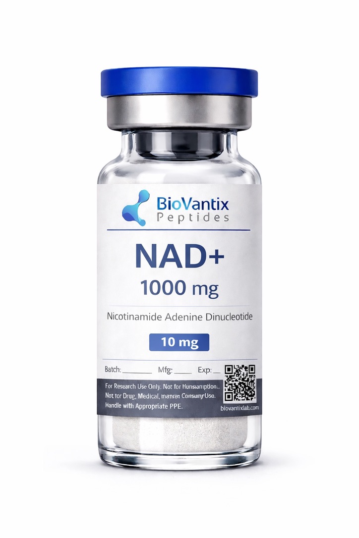
NAD+ 1000MG
NAD⁺ (nicotinamide adenine dinucleotide) is an essential cellular coenzyme involved in redox metabolism and energy production across all living systems. It functions as a critical electron carrier, cycling between its oxidized (NAD⁺) and reduced (NADH) forms to support mitochondrial oxidative phosphorylation and glycolytic energy transfer in experimental models. Beyond its metabolic role, NAD⁺ serves as a required substrate for sirtuins and poly(ADP-ribose) polymerases (PARPs), enzymes involved in DNA repair, gene expression regulation, and cellular stress responses. In preclinical research, NAD⁺ is used to investigate mitochondrial homeostasis, muscle metabolism, neuroprotection, and molecular mechanisms associated with aging and cellular resilience.
For research use only. Not for human consumption.
References:
Cantó C et al., Cell Metabolism, 2015;22(1):31–53
Verdin E., Science, 2015;350(6265):1208–1213
Yoshino J et al., Cell Metabolism, 2018;27(3):529–547
$110.00 Original price was: $110.00.$100.00Current price is: $100.00.
Out of stock
Popular Peptides
Peptide Blends
-
1. Overview
-
2. Biochemical Characteristics
-
3. Research Applications
-
4. Pathway / Mechanistic Context
-
5. Summary of Preclinical Research
-
6. Form & Analysis Testing
-
7. Referenced Citations
-
8. RUO Disclaimer
Overview
Nicotinamide adenine dinucleotide (NAD⁺) is a pyridine nucleotide cofactor that exists in both its oxidized (NAD⁺) and reduced (NADH) forms. In biochemical and cellular research, NAD⁺ is primarily studied as an electron carrier involved in redox reactions and as a substrate for multiple classes of enzymes. Published literature describes NAD⁺ as a molecule involved in intracellular metabolic networks and, in specific experimental settings, extracellular signaling measurements.
All information presented below is provided strictly within the context of preclinical research. No therapeutic, diagnostic, or medical claims are made or implied.
Biochemical Characteristics
Molecular Formula: C₂₁H₂₇N₇O₁₄P₂
Molecular Weight: 663.43 g/mol
PubChem CID: 925
CAS Number: 53-84-9
NAD⁺ functions as an active redox cofactor capable of accepting and donating electrons during enzymatic reactions. In experimental systems, NAD⁺ availability is commonly evaluated as a measurable variable associated with metabolic flux, enzymatic activity assays, and intracellular signaling. NAD⁺ is also investigated as a substrate for enzymes involved in post-translational modification processes, including ADP-ribosylation.
Research Applications
In laboratory research, NAD⁺ is used as a reference compound or experimental variable in:
- Redox biochemistry and mitochondrial metabolism assays
- Enzymatic activity measurements involving sirtuins, PARPs, and dehydrogenases
- Transcriptomic and metabolomic profiling studies reporting NAD⁺-associated expression patterns
- Cellular stress and aging model systems
- Extracellular signaling experiments measuring nucleotide release and receptor-linked responses
All applications are limited to controlled in vitro research settings or animal models.
Pathway / Mechanistic Context
Throughout the referenced literature, NAD⁺ is discussed in relation to multiple metabolic pathway–annotated datasets. These discussions are presented as reported measurements, observed associations, or differential expression trends rather than direct functional outcomes. Frequently referenced pathway contexts include:
- Redox metabolism and readouts associated with the mitochondrial electron transport chain
- Gene and protein activity measurements annotated for sirtuins
- DNA damage response datasets linked to PARP activity
- PGC-1α-associated transcriptional profiles
- Inflammation-related signaling components evaluated through cytokine endpoints or gene expression analyses
Summary of Preclinical Research
Mitochondrial and Metabolic Models
Animal and cell-based studies describe associations between NAD⁺ availability and measured mitochondrial parameters, including markers of oxidative phosphorylation and indicators of redox status. These findings are presented as dataset-level observations derived from preclinical models.
Gene Expression and Aging-Associated Datasets
Transcriptomic analyses in aging-related models report differential expression patterns across nuclear and mitochondrial gene sets under experimental conditions involving altered NAD⁺ levels. Interpretation is limited to the reported expression profiles rather than direct claims of functional restoration.
Neurodegeneration-Focused Models
In murine models of neurodegenerative disease, published studies report associations between NAD⁺ exposure and markers of neuronal survival, oxidative stress indicators, and measured mitochondrial parameters. These observations are presented as preclinical correlations within disease model systems.
Inflammation-Related Measurements
Several studies reference NAD⁺-associated datasets involving NAMPT, cytokine measurements, and inflammatory signaling components. Reported findings are based on gene expression profiles, enzymatic activity measurements, or pathway-level annotations.
Collectively, the scientific literature positions NAD⁺ as a biochemical variable used to investigate mechanisms related to metabolism, aging, and cellular stress in non-clinical research systems.
Form & Analysis Testing
This product is supplied as a research-grade compound intended exclusively for laboratory use. Analytical characterization may include chromatographic purity assessment and identity confirmation by mass spectrometry. Batch-specific specifications should be verified using the accompanying Certificate of Analysis (COA), where available.
Referenced Citations
- “NAD+ Science 101 – What Is NAD+ & Why It’s Important,” Elysium Health. [Online]. Available: https://www.elysiumhealth.com/en-us/knowledge/science-101/everything-you-need-to-know-about-nicotinamide-adenine-dinucleotide-nad. [Accessed: 25-Jul-2019].
- “Nicotinamide Riboside: Benefits, Side Effects and Dosage,” Healthline. [Online]. Available: https://www.healthline.com/nutrition/nicotinamide-riboside. [Accessed: 25-Jul-2019].
- R. T. Matthews, L. Yang, S. Browne, M. Baik, and M. F. Beal, “Coenzyme Q10 administration increases brain mitochondrial concentrations and exerts neuroprotective effects,” Proceedings of the National Academy of Sciences of the United States of America, vol. 95, no. 15, pp. 8892–8897, Jul. 1998. [PMC]
- “What You Need to Know About Resveratrol Supplements,” WebMD. [Online]. Available: https://www.webmd.com/heart-disease/resveratrol-supplements. [Accessed: 25-Jul-2019].
- N. Sun, R. J. Youle, and T. Finkel, “The Mitochondrial Basis of Aging,” Molecular Cell, vol. 61, no. 5, pp. 654–666, Mar. 2016. [PMC]
- D. Stipp, “Beyond Resveratrol: The Anti-Aging NAD Fad,” Scientific American Blog Network. [Online]. Available: https://blogs.scientificamerican.com/guest-blog/beyond-resveratrol-the-anti-aging-nad-fad/. [Accessed: 08-Jul-2019].
- A. P. Gomes et al., “Declining NAD+ Induces a Pseudohypoxic State Disrupting Nuclear-Mitochondrial Communication during Aging,” Cell, vol. 155, no. 7, pp. 1624–1638, Dec. 2013. [PMC]
- S. Imai and L. Guarente, “NAD+ and Sirtuins in Aging and Disease,” Trends in Cell Biology, vol. 24, no. 8, pp. 464–471, Aug. 2014. [PubMed]
“`
“`
- A. R. Mendelsohn and J. W. Larrick, “Partial Reversal of Skeletal Muscle Aging by Restoration of Normal NAD+ Levels,” Rejuvenation Research, vol. 17, no. 1, pp. 62–69, Feb. 2014. [PubMed]
- C. Kang, E. Chung, G. Diffee, and L. L. Ji, “Exercise Training Attenuates Aging-Associated Mitochondrial Dysfunction in Rat Skeletal Muscle: Role of PGC-1α,” Experimental Gerontology, vol. 48, no. 11, pp. 1343–1350, Nov. 2013. [PubMed]
- S. Ringholm et al., “Effect of Lifelong Resveratrol Supplementation and Exercise Training on Skeletal Muscle Oxidative Capacity in Aging Mice: Impact of PGC-1α,” Experimental Gerontology, vol. 48, no. 11, pp. 1311–1318, Nov. 2013. [PubMed]
- A. Lloret and M. F. Beal, “PGC-1α, Sirtuins and PARPs in Huntington’s Disease and Other Neurodegenerative Conditions: NAD+ to Rule Them All,” Neurochemical Research, May 2019. [PubMed]
- C. Shan et al., “Protective Effects of β-Nicotinamide Adenine Dinucleotide Against Motor Deficits and Dopaminergic Neuronal Damage in a Mouse Model of Parkinson’s Disease,” Progress in Neuro-Psychopharmacology & Biological Psychiatry, vol. 94, p. 109670, Jun. 2019. [PubMed]
- D. C. Maddison and F. Giorgini, “The Kynurenine Pathway and Neurodegenerative Disease,” Seminars in Cell & Developmental Biology, vol. 40, pp. 134–141, Apr. 2015. [PubMed]
- A. Garten, S. Schuster, M. Penke, T. Gorski, T. de Giorgis, and W. Kiess, “Physiological and Pathophysiological Roles of NAMPT and NAD Metabolism,” Nature Reviews Endocrinology, vol. 11, no. 9, pp. 535–546, Sep. 2015. [PubMed]
- S. Yamaguchi and J. Yoshino, “Adipose Tissue NAD+ Biology in Obesity and Insulin Resistance: From Mechanism to Therapy,” BioEssays, vol. 39, no. 5, May 2017. [PMC]
- J. E. Humiston, “Nicotinamide Adenine Dinucleotide,” p. 68. [FDA]
“`
“`
ALL ARTICLES AND PRODUCT INFORMATION PROVIDED ON THIS WEBSITE ARE FOR INFORMATIONAL AND EDUCATIONAL PURPOSES ONLY.
RUO Disclaimer
The products offered on this website are supplied for in vitro research only. In vitro studies (Latin: “in glass”) are performed outside the body. These products are not medicines or drugs and have not been approved by the FDA to prevent, treat, or cure any medical condition, illness, or disease. Bodily administration of any kind to humans or animals is strictly prohibited by law.
For laboratory research use only. Not for human, medical, diagnostic, or veterinary use.
| Properties | |
|---|---|
| Molecular Formula | C21H28N7O14P2+ |
| Molecular Weight | 664.4 |
| Monoisotopic Mass | 664.11694759 |
| Polar Surface Area | 318 |
| Complexity | 1130 |
| XLogP | -5.9 |
| Hydrogen Bond Acceptors | 18 |
| Rotatable Bonds | 11 |
| PubChem LCSS | Nadide (JAN/USAN/INN) Laboratory Chemical Safety Summary |
| Identifiers | |
|---|---|
| CID | 5893 |
| InChI | InChI=1S/C21H27N7O14P2/c22-17-12-19(25-7-24-17)28(8-26-12)21-16(32)14(30)11(41-21)6-39-44(36,37)42-43(34,35)38-5-10-13(29)15(31)20(40-10)27-3-1-2-9(4-27)18(23)33/h1-4,7-8,10-11,13-16,20-21,29-32H,5-6H2,(H5-,22,23,24,25,33,34,35,36,37)/p+1/t10-,11-,13-,14-,15-,16-,20-,21-/m1/s1 |
| InChIKey | BAWFJGJZGIEFAR-NNYOXOHSSA-O |
| Isomeric SMILES | C1=CC(=C[N+](=C1)[C@H]2[C@@H]([C@@H]([C@H](O2)COP(=O)(O)OP(=O)(O)OC[C@@H]3[C@H]([C@H]([C@@H](O3)N4C=NC5=C(N=CN=C54)N)O)O)O)O)C(=O)N |
| Canonical SMILES | C1=CC(=C[N+](=C1)C2C(C(C(O2)COP(=O)(O)OP(=O)(O)OCC3C(C(C(O3)N4C=NC5=C(N=CN=C54)N)O)O)O)O)C(=O)N |
| IUPAC Name | [[(2R,3S,4R,5R)-5-(6-aminopurin-9-yl)-3,4-dihydroxolan-2-yl]methoxy-hydroxyphosphoryl] [(2R,3S,4R,5R)-5-(3-carbamoylpyridin-1-ium-1-yl)-3,4-dihydroxolan-2-yl]methyl hydrogen phosphate |
Storage Instructions:
All of our products are manufactured using the lyophilization (freeze-drying) process, ensuring they remain 100% stable during shipping for up to 3–4 months.
Once peptides have been reconstituted (mixed with bacteriostatic water), they should be stored in a refrigerator to maintain stability. After reconstitution, peptides generally remain stable for up to 30 days.
Lyophilization is a specialized dehydration process, also known as freeze-drying, in which peptides are first frozen and then exposed to low pressure. This causes the water inside the peptide vial to sublime directly from a solid to a gas, leaving behind a stable, white crystalline structure known as a lyophilized peptide. This white, fluffy powder can be stored at room temperature until it is ready to be reconstituted with bacteriostatic water.
Upon receiving your peptides, it is essential to keep them cool and protected from light. If the peptides will be used immediately, or within the next few days, weeks, or months, short-term refrigeration below 4°C (39°F) is generally sufficient. Lyophilized peptides are typically stable at room temperature for several weeks or longer, so room-temperature storage is generally acceptable if they will be used within a relatively short period.
However, for long-term storage (several months to years), peptides should ideally be kept in a freezer at -80°C (-112°F). Freezing is the preferred method for preserving peptide stability over extended periods.
For more information on proper peptide storage techniques, click the following link:
http://biovantixlab.com/peptide-storage/
:::